Photodisruption in Cells using Femtosecond Laser Pulses
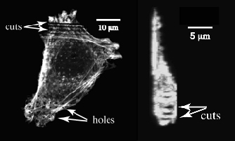
Output: Mazur and Ingber (Harvard Medical School) are studying photodisruption in cells with sub-cellular precision. The photodisruption is achieved by focusing nanojoule, femtosecond laser pulses with high numerical-aperture microscope objectives. The figure shows the central slice of a photodisrupted cell. The size of cuts through the top half of the cell, made with energy ranging from 2 nJ to 0.5 nJ is comparable to the width of a single actin fiber bundle. In the lower half of the cell, holes were punched using 100 to 500 pulses. The side view of a cell cut with 2 nJ, 100 fs laser pulses is also shown. Tissue is removed only from the middle of the cell leaving both the top and bottom layers intact.
Outcome: Our new technique allows us to photodisrupt biological tissue with great precision, with potential applications in laser microsurgery. The very small amount of energy and near-infrared wavelength used to photodisrupt sub-cellular structures makes it possible to apply this procedure to living cells for cellular structure and function research, such as the analysis of cytoskeleton integrity in living cells.
Eric Mazur, and
Ingber
Harvard MRSEC (DMR-0820484)