CARS Imaging of Cells
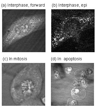
Laser-scanning CARS images of NIH3T3 fibroblasts cells. (a) Forward-detected image of a cell in interphase, tuned to 2870 cm-1 (lipid C-H stretching vibration). (b) Simultaneously epi-detected image of the same cell as in (a), which has a better contrast for small lipid vesicles. (c) Forward-detected image of a cell in metaphase, tuned to 1090 cm-1 (DNA backbone vibration). (d) Forward-detected image of cells in apoptosis, tuned to 2870 cm-1.
Xie has significantly improved the use of coherent anti-Stokes Raman scattering (CARS) for microscopy of living cells. This technique utilized vibrational spectroscopy as the contrast mechanism, concurrently allowing identification of chemical species with the imaging. The new CARS microscope utilizes simultaneous forward and epidetection of the scattering enabling fast data acquisition and high sensitivity. Xie has exploited these improvements to image cells undergoing apoptosis, or programmed death. For example, he is able to visualize the formation of lipid vesicles in the cytoplasm when the CARS microscope is tuned to the C-H stretching vibration.
Xie
Harvard MRSEC (DMR-0820484)